
We offer this service at the following locations:
Nuclear medicine procedures visualise metabolic and disease processes in the body using radioactive substances.
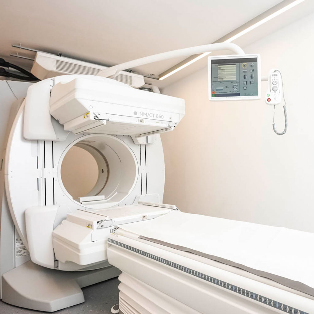
In addition to planar scintigraphy, SPECT (single photon emission tomography) is also available to assess metabolic activity in organs such as the heart, brain or bones. However, the anatomical structures of the organs can be visualised much better with computer tomography.
The latest generation SPECT/CT device, which we put into operation about a year ago, combines both examination methods and their advantages in one device and makes it possible to precisely assign the metabolic processes to the anatomical structures. This new hybrid procedure is becoming increasingly important, particularly in oncological imaging for the diagnosis and monitoring of cancer, but also for numerous orthopaedic issues.
All standard nuclear medicine examinations are offered at our centre in Munich. If you have any questions, please contact our registration desk:
Phone: +49 (0) 89 / 896 000 – 0
You will find information on preparation and information sheets in the section Preparing for the examination.
Your specialists
Dr. med. Holger Zeitler
Specialist in radiology

Dr. med. Dr. med. habil. Carola Wagner-Manslau
Specialist in nuclear medicine and radiology
Special focus
We provide a wide range of nuclear medicine examinations in our department, with a particular focus on:
- the diagnosis and treatment of diseases of the thyroid and parathyroid glands
- Skeletal scintigraphy for oncologic, orthopaedic and rheumatological issues
- Brain scintigraphy (DAT scan) in the diagnosis of Parkinson’s syndromes
- Nuclear cardiology (Myocard-scintigraphy)
- Renal function scintigraphy
- Sentinel lymph node scintigraphy
Basic information on nuclear medicine / scintigraphy at the Radiological Centre Munich
Nuclear medicine refers to the use of radioactive substances for examination and treatment purposes.
The radiopharmaceutical – the radioactive substance or the chemical compound of the radioactive substance with other substances – is normally injected into a vein in the arm. It is distributed in the body via the bloodstream and, depending on the type of substance used, accumulates in the desired organ.
For conventional nuclear medicine diagnostics, substances with a short half-life (hours to days) are usually used, which emit gamma rays and can be measured very accurately outside the body. Images of these organs can be taken with a special camera (gamma camera). The most commonly used isotope is technetium-99m (Tc-99m) with a short half-life of 6 hours.
The nuclear medicine imaging procedure, scintigraphy, primarily depicts the function of an organ or organ system, in contrast to morphological imaging procedures (X-ray, CT, MRI), which mainly show the structure.
Due to the low radiation dose in nuclear medicine examinations, there are no significant restrictions. However, examinations with radioactive substances should not be carried out during pregnancy.
Thyroid scintigraphy
- Functional assessment of the thyroid gland and thyroid nodules
- Check-up after radio-iodine therapy
Skeletal/bone scintigraphy
- Tumour aftercare
- Inflammatory bone diseases
- Loosening of joint prostheses (hip TEP, knee TEP)
- Unclear bone complaints
Myocardial scintigraphy (IGeL)
- Detection of functional disorders of the coronary arteries (coronary heart disease)
Kidney scintigraphy
- Functional disorders of the kidneys (e.g. shrunken kidneys)
- Obstruction of the ureter
- Determination of kidney function (e.g. before chemotherapy or hyperthermia)
- Unclear increase in blood pressure (exclusion of renal artery stenosis)
Pulmonary scintigraphy (IGeL)
- • Exclusion of pulmonary embolism
DAT scan (IGeL)
- • Diagnosis of Parkinson’s disease
You can also have specialised examinations carried out:
- Salivary gland scintigraphy
- Parathyroid scintigraphy (IGeL)
- Scintigraphy for the visualisation of endocrine tumours