
We offer this service at the following locations:
We are pleased to be able to offer you PET-CT as part of our range of diagnostic services with immediate effect.
PET-CT is a so-called hybrid procedure consisting of a combination of p(-ositron) e(-missions) t(-omography) and CT (computer tomography). Using a weakly radioactively labelled test substance that binds to tumours, these are made visible and can then be precisely localised anatomically on the high-resolution CT images.
PSMA (prostate-specific membrane antigen) is available to us as a prostate carcinoma-specific ligand for the diagnosis of prostate carcinomas. The labelled sugar molecule FDG (fluorodeoxy-glucose) is used for other tumours.
Examples of typical questions according to current guidelines
PSMA (Prostate-specific-membrane-antigen)
- Diagnosis of tumour spread (lymph nodes/bones) in high-risk diseases (Gleason 8 – 10, PSA > 20ng/ml, cT3 and cT4) to determine therapy
- Suspected tumour recurrence after surgery if PSA rises again
- Therapy monitoring for advanced tumour disease
- Tumour search in case of high clinical suspicion after negative multiparametric MRI and biopsy
FDG (Fluordeoxyglucose)
- for numerous tumours to determine tumour spread
- Recurrence diagnostics
- Primary tumour search
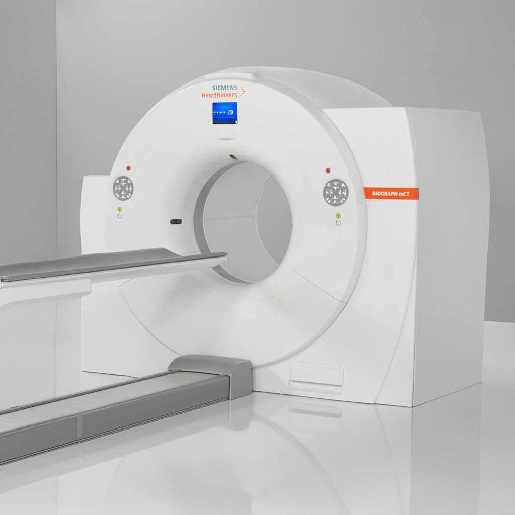
As part of an equipment pool, the examinations are carried out on a recently installed PET-CT of the latest generation (Siemens Biograph mCT) under our management at the PET-CT Munich Centre in Sonnenstr. 24. For further information, please contact us on 089/896 000 – 33.
In order to be able to plan the appointment individually, we need some information from you in advance. For prostate diagnostics, please use the following registration form PSMA PET-CT. For all other examinations, please use the following registration form (FDG). After checking the question and ensuring the availability of the respective test substance, we will arrange the exact date and time with you directly.
All examinations with PSMA and most PET-CT examinations with FDG are currently private medical services, but are regularly reimbursed by private health insurance companies.
Basic information on PET-CT
A PET-CT combines two different types of examination – in just one device. Computed tomography is used to obtain high-resolution cross-sectional images of your body. This allows us to assess the anatomy of your body and, for example, detect and categorise changes in an organ. We can also use PET (positron emission tomography) to make statements about certain metabolic processes in your tissues.
To ensure that the PET examination runs smoothly, we inject a weakly radioactive substance – usually a radioactively labelled glucose or a ligand that binds to tumour cells, such as PSMA in the case of prostate tumours – into your arm vein. The active substance is then distributed throughout your body. Many tumour cells have an increased metabolism (e.g. due to their rapid growth). The substance reacts to this. This happens because the injected active substance breaks down naturally and weak radiation is produced. This allows the distribution of the substance in the body to be visualised. By performing a CT scan at the same time, we can precisely localise these metabolic curtains in your body. The diseased areas appear coded in red on the CT images.
PET-CT enables us to answer two fundamentally different medical questions at the same time:
- What does the tissue look like?
- What are its biochemical properties?
- Are there indications of tumours or metastases?
An example:
The initial situation is a malignant cancer that fortunately responds well to chemotherapy. Long before the tumour foci finally become smaller, they show changes in their metabolism. Chemotherapy disrupts the metabolism of the tumour cells. This also reduces the uptake of the radioactively labelled substance that we inject into you for the PET scan. This means that we can tell you much earlier whether the chemotherapy you are currently receiving is right for you – or whether the treatment regimen should be changed.
Firstly, we give you an injection into the vein in your arm, which contains the radioactive substance. So that this so-called tracer can be distributed in your body via the cell metabolism, you will then have a rest period of around 1.5 hours during which you should move as little as possible. A specially equipped relaxation room is available for this purpose.
The PET-CT will then begin. You will lie relaxed on a couch that is automatically pushed through the PET-CT machine. The machine has a large opening like a CT scanner, so that claustrophobic patients usually have no problems. You will receive breathing instructions via a loudspeaker. Please maintain your lying position until the end of the examination. This is the only way to obtain meaningful images. During the examination, which takes about 30 to 45 minutes, we will always be close to you and keep an eye on you.
Afterwards, we will take a close look at the images so that we can inform you of the results.